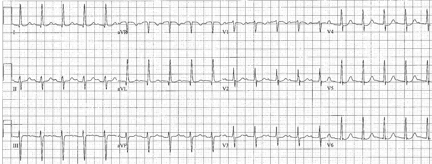
CMC ECG MASTERS
  • Home
    • About Us
    • EM GuideWire
    • CMC Ed Masters
    • Ped EM Morsels
  • Basics
  • P Waves
    • Sinus
    • Not sinus
    • Absent
  • PR interval
    • Short PR
    • Prolonged PR
    • Variable PR
  • QRS
    • Wide
    • Axis >
      • LAD
      • RAD
    • Amplitude >
      • High Voltage
      • Low Voltage
      • Alternans
    • R Wave Progression
    • Pathological Q waves
  • ST Segments
    • ST Elevation
    • ST Depression
  • T Waves
    • Flat, Bifid, or Notched
    • Inverted
  • QT intervals
    • Prolonged QT
    • Short QT
  • Patterns
    • STEMI
    • Pulmonary Embolism
    • Ventricular Hypertrophy
    • Pulmonary Disease
    • ST-T Patterns
    • Electrolyte Abnormalities
    • Cases >
      • Case 1
      • Case 2
      • Case 3
      • Case 4
      • Case 5
      • Case 6
      • Case 7
      • Case 8
      • Case 9
      • Case 10
      • Case 11
      • Case 12
      • Case 13
      • Case 14
      • Case 15
      • Case 16
      • Case 17
      • Case 18
      • Case 19
  • Interpreter

Left Axis Deviation

Causes of left axis deviation
  • Advanced age
  • LVH
  • Obesity
  • Emphysema
  • Inferior MI (loss of inferior forces)
  • Left anterior fascicular block (LAFB); diagnostic criteria:
    • left axis deviation > - 45 degrees
    • leads I and aVL ­ but start with a narrow Q (qR)
    • leads II, III, aVF  but start with a small r (rS)
    • QRS may be slightly widened but < 0.12s
Picture
Wide QRS
RAD
Amplitude
  • Home
    • About Us
    • EM GuideWire
    • CMC Ed Masters
    • Ped EM Morsels
  • Basics
  • P Waves
    • Sinus
    • Not sinus
    • Absent
  • PR interval
    • Short PR
    • Prolonged PR
    • Variable PR
  • QRS
    • Wide
    • Axis >
      • LAD
      • RAD
    • Amplitude >
      • High Voltage
      • Low Voltage
      • Alternans
    • R Wave Progression
    • Pathological Q waves
  • ST Segments
    • ST Elevation
    • ST Depression
  • T Waves
    • Flat, Bifid, or Notched
    • Inverted
  • QT intervals
    • Prolonged QT
    • Short QT
  • Patterns
    • STEMI
    • Pulmonary Embolism
    • Ventricular Hypertrophy
    • Pulmonary Disease
    • ST-T Patterns
    • Electrolyte Abnormalities
    • Cases >
      • Case 1
      • Case 2
      • Case 3
      • Case 4
      • Case 5
      • Case 6
      • Case 7
      • Case 8
      • Case 9
      • Case 10
      • Case 11
      • Case 12
      • Case 13
      • Case 14
      • Case 15
      • Case 16
      • Case 17
      • Case 18
      • Case 19
  • Interpreter