CMC ECG MASTERS
  • Home
    • About Us
    • EM GuideWire
    • CMC Ed Masters
    • Ped EM Morsels
  • Basics
  • P Waves
    • Sinus
    • Not sinus
    • Absent
  • PR interval
    • Short PR
    • Prolonged PR
    • Variable PR
  • QRS
    • Wide
    • Axis >
      • LAD
      • RAD
    • Amplitude >
      • High Voltage
      • Low Voltage
      • Alternans
    • R Wave Progression
    • Pathological Q waves
  • ST Segments
    • ST Elevation
    • ST Depression
  • T Waves
    • Flat, Bifid, or Notched
    • Inverted
  • QT intervals
    • Prolonged QT
    • Short QT
  • Patterns
    • STEMI
    • Pulmonary Embolism
    • Ventricular Hypertrophy
    • Pulmonary Disease
    • ST-T Patterns
    • Electrolyte Abnormalities
    • Cases >
      • Case 1
      • Case 2
      • Case 3
      • Case 4
      • Case 5
      • Case 6
      • Case 7
      • Case 8
      • Case 9
      • Case 10
      • Case 11
      • Case 12
      • Case 13
      • Case 14
      • Case 15
      • Case 16
      • Case 17
      • Case 18
      • Case 19
  • Interpreter

ST Segment Elevation

CAUSES OF ST ELEVATION
Ischemia, injury, infarction (i - i - i )
Characteristics: chest pain; ST­ elevation localized to a wall/vascular distribution; Q waves
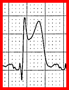
  • Acute or recent MI:
    • Hyperacute: no Q, T elevation
    • Subacute: R downgoing, T downgoing
    • Later: pathologic Qs
  • Prinzmetal angina:  like hyperacute MI but transient; no associated Q or T wave changes
  • Cardiac trauma: ​​(contusion, stab wound, GSW): persistent ST but no progression of T, Q
  • Subacute cardiac rupture:  progressive ST elevation in Q wave leads
  • ST elevation in aVR with diffuse ST depression elsewhere:
    • possible left main obstruction
  • Myocarditis
  • Following high-energy cardioversion
  • Acute adrenergic stress: pressors, inotropes, emotional stress; frequently present in ICU
  • LV aneurysm: persistent ST elevation in Q wave leads
Picture
Hyperacute STE
Picture
STEMI

SECONDARY ST ELEVATION
Characteristics: wide QRS complexes or LVH; mirror image of ST depression
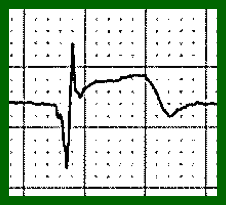
  • LBBB: coved, elevated ST-Ts in leads with downgoing QRS complexes
  • WPW: as above
  • Paced rhythm: as above
  • LVH: upward concave ST elevation and upright Ts in V1-V2 (mirror image strain)
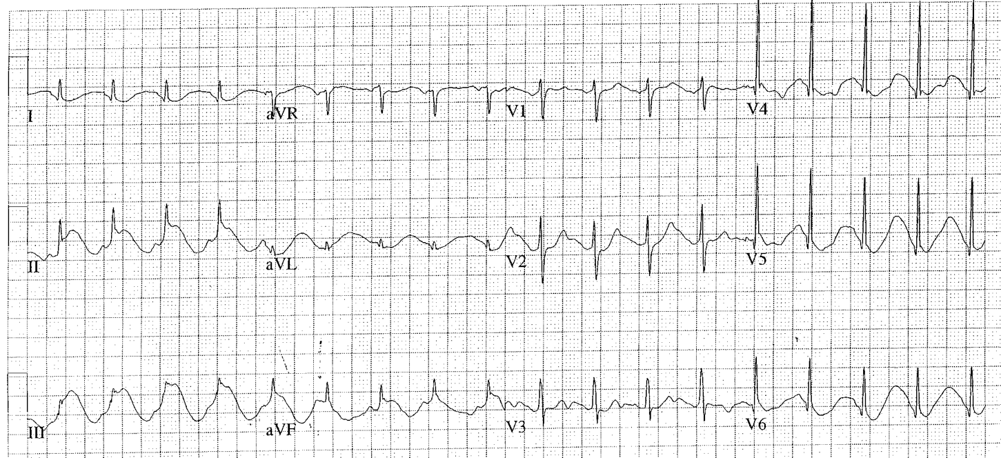
Picture
LBBB

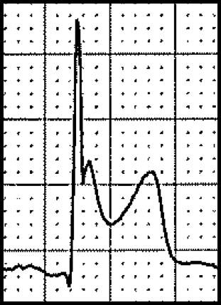
TERMINAL NOTCHING OF THE QRS COMPLEX FOLLOWED BY HAMMOCK-SHAPED ST ELEVATION
  • ​Heart rate fast - pericarditis: diffuse ST elevation; depressed PR segments (esp. in II)
  • HR normal - early repolarization: triphasic QRS with terminal notch; upright Ts; normal QT
  • HR slow - hypothermia: Osborn wave; prolonged QT; shivering artifact
  • Early repolarization variant: early repolarization-type ST elevation followed by inverted Ts – usually seen in AA males with LVH, and with acute or chronic cocaine use

ELECTROLYTE ABNORMALITIES/DRUG EFFECTS
  • ​Hypercalcemia: coved ST elevation and absence of Ts in anteroseptal leads (STs are probably Ts)
  • Digitalis: scooped ST elevation in anteroseptal leads; mirror image of scooped ST in lateral leads
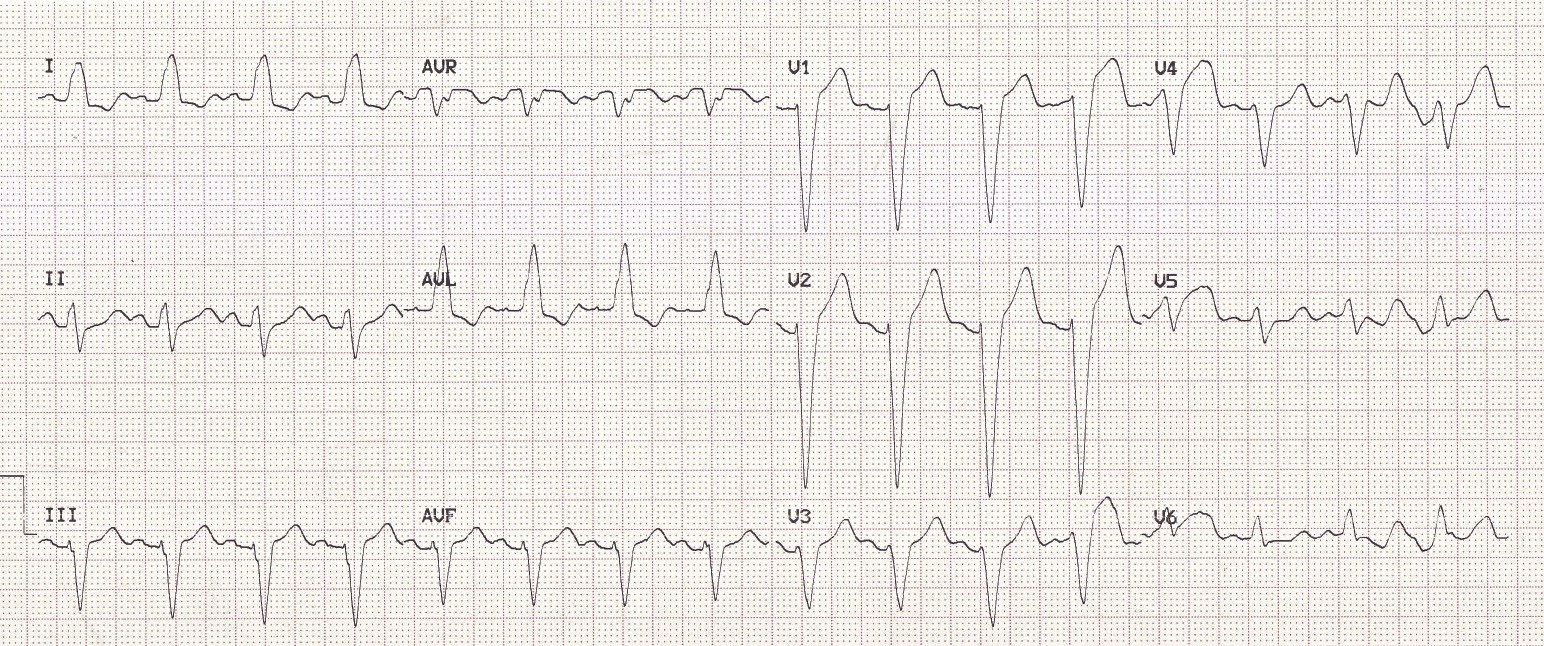
  • Hyperkalemia: ST elevation in anteroseptal leads; narrow-based peaked Ts; “Brugada pattern”
  • Na-channel blocker toxicity: (including TCA, cocaine): “Brugada pattern”
Picture
Hypercalcemia

MISCELLANEOUS
  • ​Acute CNS disorder: (SAH, ICH, trauma): QT frequently prolonged
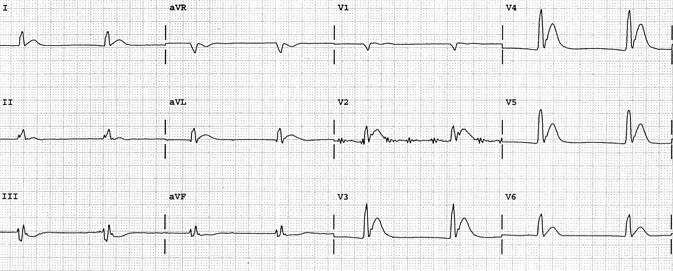
  • Brugada syndrome: V1-2: rSR’, coved ST elevation, T (high take-off ST elevation followed by T downgoing)
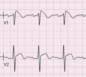
Picture
Brugada

PSEUDO-ST ELEVATION
  • Atrial flutter: regular SVT at ~150/min; flutter waves may mimic ST elevation
  • Artifact: ST changes from cycle to cycle (ST elevation does not respect the cardiac cycle)
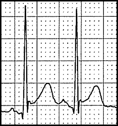
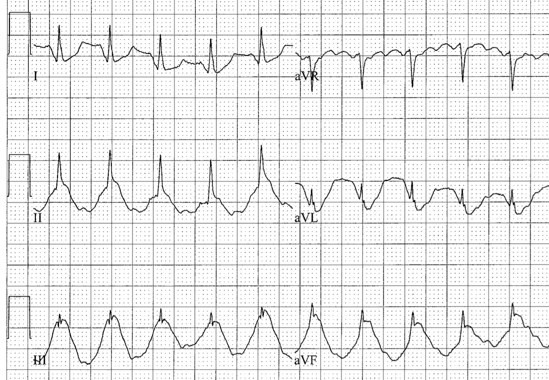
Picture

SPIKED HELMET SIGN
  • Does respect the cardiac cycle: most likely physiologic signal (repetitive epidermal stretch from increased intracavitary pressure)
  • Suggests acute intrathoracic or intraabdominal event
  • High risk condition, high hospital mortality
  • Recognition of the spiked helmet sign should lead you to search for acute pathology
    • If present in the inferior leads, consider acute abdominal event
    • If present in the chest leads, consider acute thoracic event
  • Prompt recognition may help find and treat the acute non-cardiac condition
Picture
Picture
Back to QRS
ST Depression
T Waves
  • Home
    • About Us
    • EM GuideWire
    • CMC Ed Masters
    • Ped EM Morsels
  • Basics
  • P Waves
    • Sinus
    • Not sinus
    • Absent
  • PR interval
    • Short PR
    • Prolonged PR
    • Variable PR
  • QRS
    • Wide
    • Axis >
      • LAD
      • RAD
    • Amplitude >
      • High Voltage
      • Low Voltage
      • Alternans
    • R Wave Progression
    • Pathological Q waves
  • ST Segments
    • ST Elevation
    • ST Depression
  • T Waves
    • Flat, Bifid, or Notched
    • Inverted
  • QT intervals
    • Prolonged QT
    • Short QT
  • Patterns
    • STEMI
    • Pulmonary Embolism
    • Ventricular Hypertrophy
    • Pulmonary Disease
    • ST-T Patterns
    • Electrolyte Abnormalities
    • Cases >
      • Case 1
      • Case 2
      • Case 3
      • Case 4
      • Case 5
      • Case 6
      • Case 7
      • Case 8
      • Case 9
      • Case 10
      • Case 11
      • Case 12
      • Case 13
      • Case 14
      • Case 15
      • Case 16
      • Case 17
      • Case 18
      • Case 19
  • Interpreter