CMC ECG MASTERS
  • Home
    • About Us
    • EM GuideWire
    • CMC Ed Masters
    • Ped EM Morsels
  • Basics
  • P Waves
    • Sinus
    • Not sinus
    • Absent
  • PR interval
    • Short PR
    • Prolonged PR
    • Variable PR
  • QRS
    • Wide
    • Axis >
      • LAD
      • RAD
    • Amplitude >
      • High Voltage
      • Low Voltage
      • Alternans
    • R Wave Progression
    • Pathological Q waves
  • ST Segments
    • ST Elevation
    • ST Depression
  • T Waves
    • Flat, Bifid, or Notched
    • Inverted
  • QT intervals
    • Prolonged QT
    • Short QT
  • Patterns
    • STEMI
    • Pulmonary Embolism
    • Ventricular Hypertrophy
    • Pulmonary Disease
    • ST-T Patterns
    • Electrolyte Abnormalities
    • Cases >
      • Case 1
      • Case 2
      • Case 3
      • Case 4
      • Case 5
      • Case 6
      • Case 7
      • Case 8
      • Case 9
      • Case 10
      • Case 11
      • Case 12
      • Case 13
      • Case 14
      • Case 15
      • Case 16
      • Case 17
      • Case 18
      • Case 19
  • Interpreter

Sinus P WAVES

Now that you know it is sinus, look for any abnormal Sinus P waves

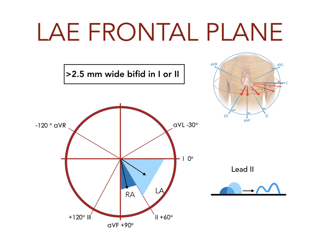
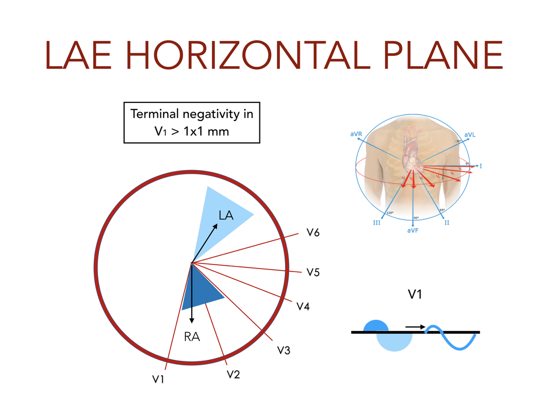
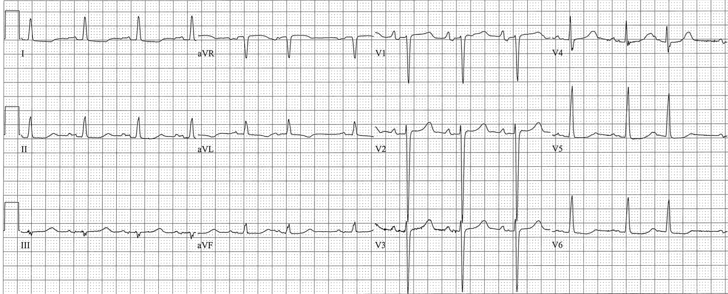
Left Atrial Enlargement

Picture
Picture
Features:
  • V1: Terminal Negativity > 1mm (sensitive, not specific)
  • Lead I or II: P wave is wide (>2.5 mm) and bifid (specific, not sensitive) 
  • 1st degree AV block (PR interval > 5 little boxes, 0.2sec)
  • PR segment Depression in II

 Clinical Significance of LAE

Findings on of LAE on EKG may suggest​
  • True left atrial enlargement
  • Left atrial hypertrophy
  • Left atrial scarring
  • Biatrial abnormality
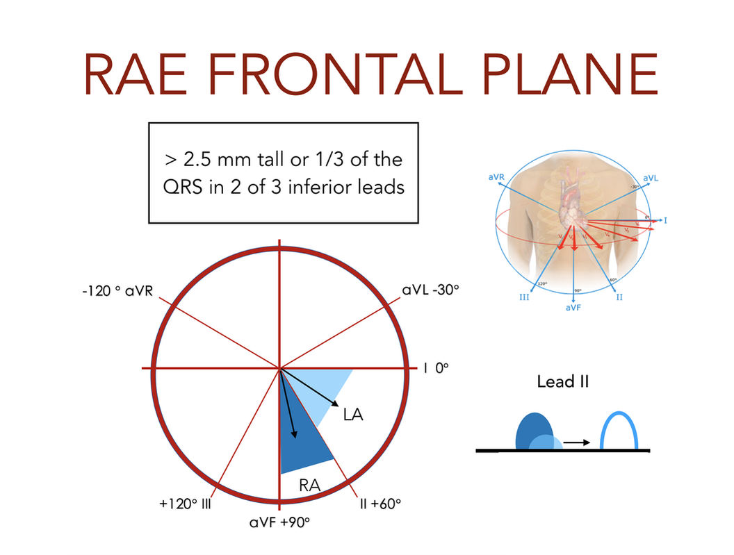
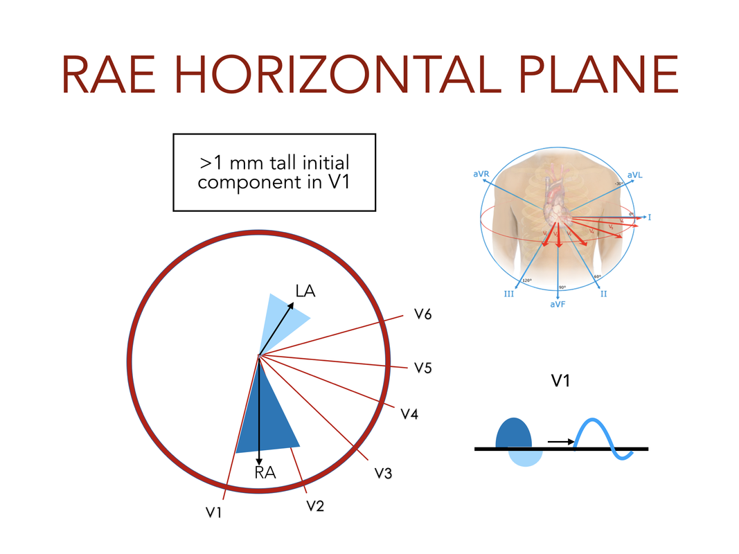
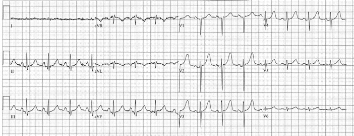
These, and frequent PACs, are risk factors for atrial fibrillation

R​IGHT ATRIAL ENLARGEMENT

Picture
Picture
Features
  • V1: initial upgoing component > 1mm (specific, not sensitive)
  • II, III, aVF: Tall and peaked P (2.5mm or 1/3 QRS height in 2 of 3 (sensitive, not specific)
  • Flat line in lead I, and inverted P, QRS in aVL (strongly suggests emphysema)​

Clinical Significance of RAE

  • RAE suggests pulmonary disease or right heart failure (especially with Reactive Airways)
  • Flatline in lead 1 and negative P and QRS in aVL suggests emphysema (vertical lie of the heart)​
  • RAE is a risk factor for Atrial Flutter
ECG has limited specificity for RAE:
  • Sinus Tachycardia is frequently accompanied by tall peaked Ps
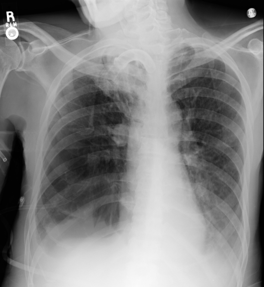
Picture
Picture
Emphysema
Picture
Biatrial enlargement
Not Sinus
Absent P Waves
Move on to PR Interval
Back
  • Home
    • About Us
    • EM GuideWire
    • CMC Ed Masters
    • Ped EM Morsels
  • Basics
  • P Waves
    • Sinus
    • Not sinus
    • Absent
  • PR interval
    • Short PR
    • Prolonged PR
    • Variable PR
  • QRS
    • Wide
    • Axis >
      • LAD
      • RAD
    • Amplitude >
      • High Voltage
      • Low Voltage
      • Alternans
    • R Wave Progression
    • Pathological Q waves
  • ST Segments
    • ST Elevation
    • ST Depression
  • T Waves
    • Flat, Bifid, or Notched
    • Inverted
  • QT intervals
    • Prolonged QT
    • Short QT
  • Patterns
    • STEMI
    • Pulmonary Embolism
    • Ventricular Hypertrophy
    • Pulmonary Disease
    • ST-T Patterns
    • Electrolyte Abnormalities
    • Cases >
      • Case 1
      • Case 2
      • Case 3
      • Case 4
      • Case 5
      • Case 6
      • Case 7
      • Case 8
      • Case 9
      • Case 10
      • Case 11
      • Case 12
      • Case 13
      • Case 14
      • Case 15
      • Case 16
      • Case 17
      • Case 18
      • Case 19
  • Interpreter