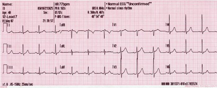
CMC ECG MASTERS
  • Home
    • About Us
    • EM GuideWire
    • CMC Ed Masters
    • Ped EM Morsels
  • Basics
  • P Waves
    • Sinus
    • Not sinus
    • Absent
  • PR interval
    • Short PR
    • Prolonged PR
    • Variable PR
  • QRS
    • Wide
    • Axis >
      • LAD
      • RAD
    • Amplitude >
      • High Voltage
      • Low Voltage
      • Alternans
    • R Wave Progression
    • Pathological Q waves
  • ST Segments
    • ST Elevation
    • ST Depression
  • T Waves
    • Flat, Bifid, or Notched
    • Inverted
  • QT intervals
    • Prolonged QT
    • Short QT
  • Patterns
    • STEMI
    • Pulmonary Embolism
    • Ventricular Hypertrophy
    • Pulmonary Disease
    • ST-T Patterns
    • Electrolyte Abnormalities
    • Cases >
      • Case 1
      • Case 2
      • Case 3
      • Case 4
      • Case 5
      • Case 6
      • Case 7
      • Case 8
      • Case 9
      • Case 10
      • Case 11
      • Case 12
      • Case 13
      • Case 14
      • Case 15
      • Case 16
      • Case 17
      • Case 18
      • Case 19
  • Interpreter

R Wave Progression

Normal QRS progression in chest leads
  • QRS predominantly downgoing in V1
  • QRS predominantly upgoing in V5 or V6
  • There is a gradual increase in the R/S ratio from V1 -> V4
Picture
QRS predominantly upgoing in V1 (R/S ratio > 1) – differential diagnosis:
  • QRS wide>0.12
    • ​RBBB: V1 rSR'
    • VT: fast, V1 not consistent with RBBB, typically no P-QRS relationship
    • WPW: short PR, delta wave, V1 not consistent with RBBB
    • BiV paced: pacer spikes in front of QRS complexes
  • QRS narrow <.11​​
    • ​RVH: T negative in V1, RAD, deep S waves in V5-V6, clinical picture (emphysema)
    • Posterior MI: T upright in V1, inverted Ts in lateral and inferior leads, clinical picture (chest pain)
    • Subtle preexcitation: short to short-normal PR, subtle delta wave
    • V1-V3 lead reversal: R wave regression from V1 to V3, may be read as anterior MI, biphasic P wave in V3
    • Normal Variant: no other signs of WPW, RVH, MI
QRS predominantly downgoing in V5 and V6
  • ​QRS wide>0.12
    • paced rhythm
    • WPW
    • VT
  • QRS narrow <.11
    • ​emphysema
    • RVH
    • LAFB
    • lateral MI
Regression of R/S ratio in chest leads
  • Possible anterior MI
  • Severe emphysema, RVH
  • Morbid obesity
  • RBBB
  • Dextrocardia
  • Incorrect electrode placement
Amplitude
Pathologic Q Waves
ST Segments
  • Home
    • About Us
    • EM GuideWire
    • CMC Ed Masters
    • Ped EM Morsels
  • Basics
  • P Waves
    • Sinus
    • Not sinus
    • Absent
  • PR interval
    • Short PR
    • Prolonged PR
    • Variable PR
  • QRS
    • Wide
    • Axis >
      • LAD
      • RAD
    • Amplitude >
      • High Voltage
      • Low Voltage
      • Alternans
    • R Wave Progression
    • Pathological Q waves
  • ST Segments
    • ST Elevation
    • ST Depression
  • T Waves
    • Flat, Bifid, or Notched
    • Inverted
  • QT intervals
    • Prolonged QT
    • Short QT
  • Patterns
    • STEMI
    • Pulmonary Embolism
    • Ventricular Hypertrophy
    • Pulmonary Disease
    • ST-T Patterns
    • Electrolyte Abnormalities
    • Cases >
      • Case 1
      • Case 2
      • Case 3
      • Case 4
      • Case 5
      • Case 6
      • Case 7
      • Case 8
      • Case 9
      • Case 10
      • Case 11
      • Case 12
      • Case 13
      • Case 14
      • Case 15
      • Case 16
      • Case 17
      • Case 18
      • Case 19
  • Interpreter