CMC ECG MASTERS
  • Home
    • About Us
    • EM GuideWire
    • CMC Ed Masters
    • Ped EM Morsels
  • Basics
  • P Waves
    • Sinus
    • Not sinus
    • Absent
  • PR interval
    • Short PR
    • Prolonged PR
    • Variable PR
  • QRS
    • Wide
    • Axis >
      • LAD
      • RAD
    • Amplitude >
      • High Voltage
      • Low Voltage
      • Alternans
    • R Wave Progression
    • Pathological Q waves
  • ST Segments
    • ST Elevation
    • ST Depression
  • T Waves
    • Flat, Bifid, or Notched
    • Inverted
  • QT intervals
    • Prolonged QT
    • Short QT
  • Patterns
    • STEMI
    • Pulmonary Embolism
    • Ventricular Hypertrophy
    • Pulmonary Disease
    • ST-T Patterns
    • Electrolyte Abnormalities
    • Cases >
      • Case 1
      • Case 2
      • Case 3
      • Case 4
      • Case 5
      • Case 6
      • Case 7
      • Case 8
      • Case 9
      • Case 10
      • Case 11
      • Case 12
      • Case 13
      • Case 14
      • Case 15
      • Case 16
      • Case 17
      • Case 18
      • Case 19
  • Interpreter

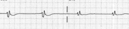
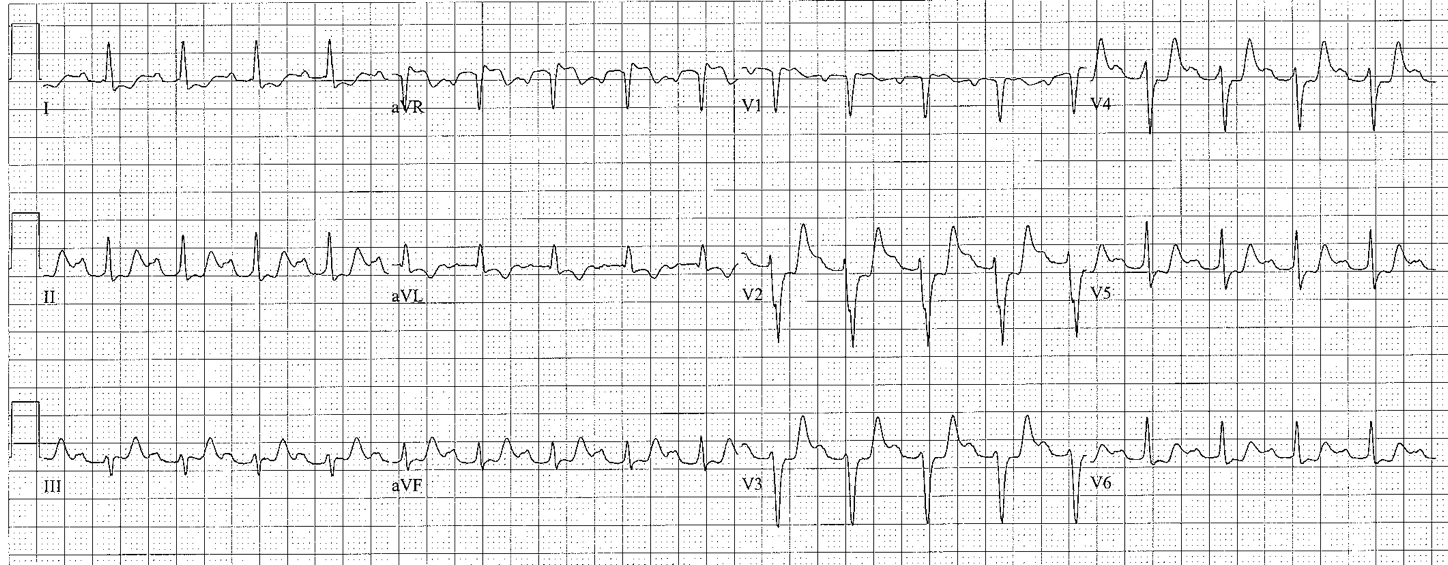
ST Segment Depression

Horizontal or downsloping ST with upright T waves-> probable ischemia
  • “Someone stepped on the ST segment”
  • May be diffuse or localized
  • Diffuse ST depression with ST elevation in aVR: possible left main obstruction
  • ST depression during PSVT is not diagnostic
Picture

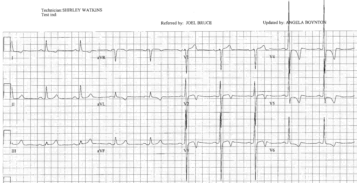
Downsloping ST depression with inverted T waves -> probable LV strain​
  • Usually in association with LVH
  • LVH, strain and chest pain: ECG not very useful
Picture

Upsloping ST depression (J point depression) -> nonspecific
  • ST usually back to baseline 2 mm after the end of QRS
  • Causes: anemia, metabolic abnormalities, MVP, normal variant

Scooped ST depression -> digitalis or hypercalcemia
  • Usually seen in left leads (I, V5-V6)
  • QT interval may be shortened
  • May be associated with other markers of digitalis effect
    • Bradycardia, first degree AV block, atrial fibrillation with slow
    • Ventricular response, digitalis-toxic tachyarrhythmias
Picture
Back to QRS
ST Elevation
T Waves
  • Home
    • About Us
    • EM GuideWire
    • CMC Ed Masters
    • Ped EM Morsels
  • Basics
  • P Waves
    • Sinus
    • Not sinus
    • Absent
  • PR interval
    • Short PR
    • Prolonged PR
    • Variable PR
  • QRS
    • Wide
    • Axis >
      • LAD
      • RAD
    • Amplitude >
      • High Voltage
      • Low Voltage
      • Alternans
    • R Wave Progression
    • Pathological Q waves
  • ST Segments
    • ST Elevation
    • ST Depression
  • T Waves
    • Flat, Bifid, or Notched
    • Inverted
  • QT intervals
    • Prolonged QT
    • Short QT
  • Patterns
    • STEMI
    • Pulmonary Embolism
    • Ventricular Hypertrophy
    • Pulmonary Disease
    • ST-T Patterns
    • Electrolyte Abnormalities
    • Cases >
      • Case 1
      • Case 2
      • Case 3
      • Case 4
      • Case 5
      • Case 6
      • Case 7
      • Case 8
      • Case 9
      • Case 10
      • Case 11
      • Case 12
      • Case 13
      • Case 14
      • Case 15
      • Case 16
      • Case 17
      • Case 18
      • Case 19
  • Interpreter