
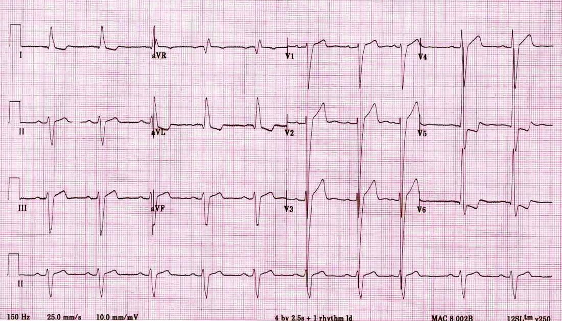
Ventricular Hypertrophy
LVH with strain
- Voltage criteria for LVH
- Left atrial enlargement
- Left axis deviation
- Subtle QRS widening (0.11 - 0.13 s)
- Repolarization abnormality (strain pattern or any other ST-T abnormality)
- Definition of strain pattern: “Upward convex ST depression followed by non-symmetrical T wave inversion (shallow downslope, rapid upslope) in leads with upright QRS complexes”
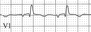
- “Mirror image” strain pattern may cause ST elevation in V1-V2
- ECG evaluation for “coronary ischemia” is limited in the presence of LVH with strain
|
RVH
|
|