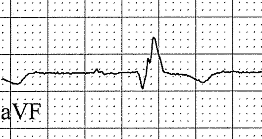
Pathological q Waves
Width: ≥ 0.03s
Depth: ≥ ⅓ of QRS amplitude
Needs to be present in at least 2 neighboring leads
Depth: ≥ ⅓ of QRS amplitude
Needs to be present in at least 2 neighboring leads
MYOCARDIAL INFARCTION
ECG Leads |
Location of MI |
Probable Culprit |
II, III, aVF (±V5,V6) |
Inferior |
RCA (or dominant LCX) |
Mirror image V1-V2 (R, ST¯, T) |
Posterolateral |
LCX |
II-III-aVF, plus V1 and RV4 |
Inferior + RV |
Proximal RCA |
V1-V4 |
Anteroseptal |
LAD |
V1-V6 (± I, aVL) |
Extensive Anterior |
LAD |
I, aVL, V4-V6 |
Lateral |
LCX |
I, aVL, V2 (± mirror image III) |
High Lateral |
LAD-D1 |
NONINFARCTION Q WAVES
- LBBB
- WPW
- LVH
- Hyperkalemia
CLINICAL SIGNIFICANCE
REMOTE MI WITH NONSPECIFIC IVCD
|
Coexisting considerations:
|

