CMC ECG MASTERS
  • Home
    • About Us
    • EM GuideWire
    • CMC Ed Masters
    • Ped EM Morsels
  • Basics
  • P Waves
    • Sinus
    • Not sinus
    • Absent
  • PR interval
    • Short PR
    • Prolonged PR
    • Variable PR
  • QRS
    • Wide
    • Axis >
      • LAD
      • RAD
    • Amplitude >
      • High Voltage
      • Low Voltage
      • Alternans
    • R Wave Progression
    • Pathological Q waves
  • ST Segments
    • ST Elevation
    • ST Depression
  • T Waves
    • Flat, Bifid, or Notched
    • Inverted
  • QT intervals
    • Prolonged QT
    • Short QT
  • Patterns
    • STEMI
    • Pulmonary Embolism
    • Ventricular Hypertrophy
    • Pulmonary Disease
    • ST-T Patterns
    • Electrolyte Abnormalities
    • Cases >
      • Case 1
      • Case 2
      • Case 3
      • Case 4
      • Case 5
      • Case 6
      • Case 7
      • Case 8
      • Case 9
      • Case 10
      • Case 11
      • Case 12
      • Case 13
      • Case 14
      • Case 15
      • Case 16
      • Case 17
      • Case 18
      • Case 19
  • Interpreter

ST-t Patterns

T-QT Pattern (not an established name)
  • Large, sometimes global T wave inversion
  • T waves may be giant negative; occasionally giant positive
  • Prolonged QT
  • A stereotypical response to a variety of noxious stimuli
  • Usually evolves ~24 hrs after the insult
  • Causes:
    • Acute CNS disorders (SAH, CVA, thalamic stroke, brain tumor, status epilepticus)
    • Catecholamine effect (-adrenergic agonist inotropes, inhalers, cocaine, pheochromocytoma)
    • Emotional stress (Tako-Tsubo cardiomyopathy)
    • Pulmonary edema
    • Massive PE

Clinical Significance

  • Most likely common cause: acute adrenergic insult
  • ECG evolution: typically evolves “next day”; gradual slow resolution in days or weeks
  • Female > male
  • Troponin elevation, LV dysfunction common
  • Prognosis is benign
  • Treatment: underlying condition; b-blocker (except for the asthmatic)
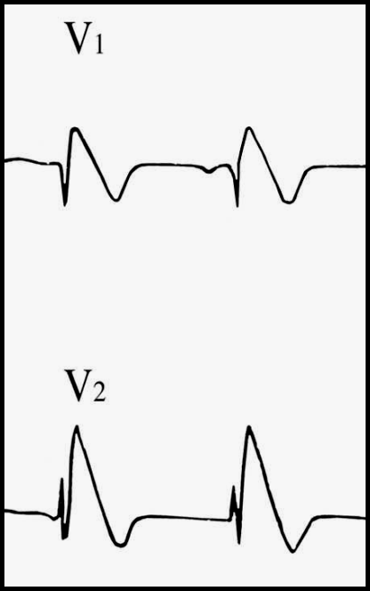
Brugada Pattern
  • RBBB pattern in V1 (rSR’: terminal positivity of QRS complex but QRS < 120 ms)
  • High take-off coved (upward convex) ST elevation in V1 and V2
  • Inverted T waves in V1 and V2
  • Causes:
    • Brugada syndrome: No underlying structural heart disease; unexplained VF/arrest; endemic in Southeast Asia; familial-sporadic otherwise; except for V1-V2, ECG is usually normal
    • Possible Brugada syndrome: no underlying structural heart disease; no h/o VF/arrest but either a family h/o unexplained sudden death or a personal h/o unexplained syncope; Southeast Asian male; except for V1-V2, the ECG is usually normal
    • Normal variant: none of the above risk factors (incidental finding); except for V1-V2, the ECG is usually normal
    • Severe hyperkalemia: Brugada pattern is associated with very wide QRS, abnormal QRS axis
    • Sodium-channel blocker toxicity (including cocaine, TCA): Brugada pattern is associated with very wide QRS, abnormal QRS axis
    • Propofol infusion syndrome (with high-dose propofol): QRS narrow; acidosis, rhabdo, ARF, hyperkalemia, hypertriglyceridemia, high risk of VT-VF and sudden death
    • Right ventricular pathology or injury
Picture

Clinical Significance

  • The only treatment that can prevent SCD is implantation of an ICD
  • The majority of patients with the Brugada ECG are not at a high risk for sudden cardiac death
  • In critically ill ICU patient: consider hyperkalemia
  • If patient was “found down”: consider sodium channel blocker toxicity
  • Risk stratification is crucial
    • Resuscitated sudden death (high risk)
    • Documented VT
    • Personal h/o unexplained syncope
    • Family h/o unexplained sudden death at a young age
    • Southeast Asian ethnicity
    • Asymptomatic (low risk)

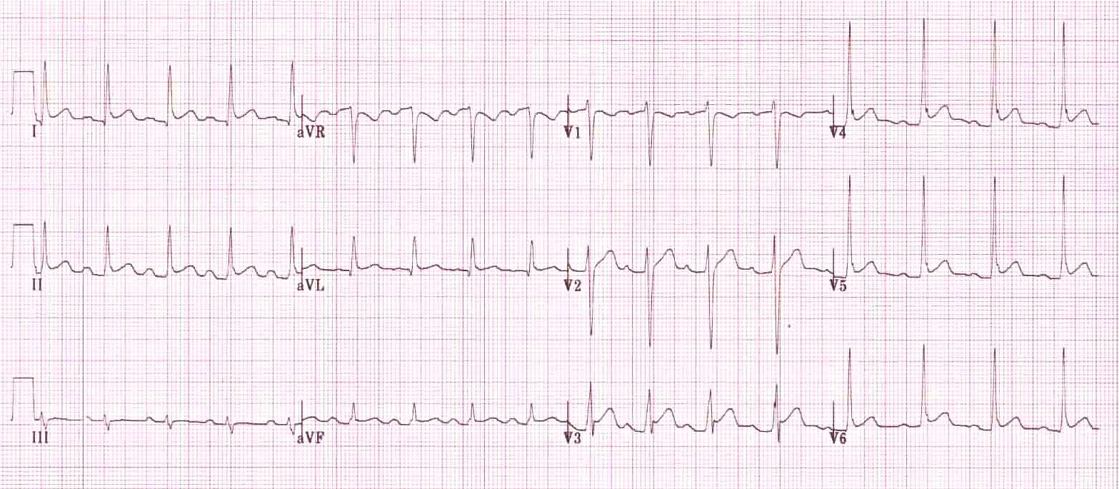
Terminal positivity of QRS (terminal notch and hammock-shaped ST elevation)
  • “Early repolarization” (normal variant)
    • Best seen in V4
    • QRS triphasic (up, down, up again)
    • Upward concave ST elevation starts from the upsloping QRS (this may cause a notch)
    • Normal, upright T waves
    • QT is normal
  • Hypothermia
    • Terminal QRS notch more prominent (Osborn wave or J wave)
    • Frequently associated with sinus bradycardia or slow atrial fibrillation
    • Marked ST-T abnormalities may be present (both ST and ST)
    • Prolonged QT
    • When rewarming: shivering artifact
  • Pericarditis
    • Diffuse ST elevation; ST elevation usually spares aVR and V1
    • Usually associated with sinus tachycardia
    • P-R segments may be depressed (especially in II) or elevated in aVR
Picture
Pulmonary Disease
Electrolyte Abnormalities
  • Home
    • About Us
    • EM GuideWire
    • CMC Ed Masters
    • Ped EM Morsels
  • Basics
  • P Waves
    • Sinus
    • Not sinus
    • Absent
  • PR interval
    • Short PR
    • Prolonged PR
    • Variable PR
  • QRS
    • Wide
    • Axis >
      • LAD
      • RAD
    • Amplitude >
      • High Voltage
      • Low Voltage
      • Alternans
    • R Wave Progression
    • Pathological Q waves
  • ST Segments
    • ST Elevation
    • ST Depression
  • T Waves
    • Flat, Bifid, or Notched
    • Inverted
  • QT intervals
    • Prolonged QT
    • Short QT
  • Patterns
    • STEMI
    • Pulmonary Embolism
    • Ventricular Hypertrophy
    • Pulmonary Disease
    • ST-T Patterns
    • Electrolyte Abnormalities
    • Cases >
      • Case 1
      • Case 2
      • Case 3
      • Case 4
      • Case 5
      • Case 6
      • Case 7
      • Case 8
      • Case 9
      • Case 10
      • Case 11
      • Case 12
      • Case 13
      • Case 14
      • Case 15
      • Case 16
      • Case 17
      • Case 18
      • Case 19
  • Interpreter